Preventive Vascular Medicine
Preventive Vascular Medicine
Vascular Health
Detection of Vascular Diseases involving veins and Peripheral Arteries remains most under diagnosed and Under treated. Around 80% of patients require only medications and don’t have the tools that they need to alert them with early warning signs before the critical symptoms.
The vascular system is the body’s essential network of Blood vessels, extending its network through Arteries and veins, catering to healthy working of Heart, Brain, kidneys, and Peripheries (Upper limbs and lower limbs). Diseases compromising the function of the network of arteries and veins is mostly widespread, but many patients are unaware of the underlying risks and common signs and symptoms. The underlying pathogenesis of Peripheral Arterial Disease is consistent with that of coronary artery disease, and almost one-fourth (25%) of the patients with Coronary Artery Disease had a concomitant Peripheral Arterial Disease.
Early vascular evaluation can’significantly supplement’ in identification of status of Vascular health in patients over 50 years of age, monitor the progression in patients who are on Medical therapy, ultimately saving lives and preventing amputations, eliminating resource wastage.
Vascular health can normally deteriorate with aging and in anyone with risk factors such as diabetes, high blood pressure, elevated cholesterol levels, and smoking.
The objectives of these Vascular evaluation can assist in examining adherence to medical therapy among a cohort of patients treated with medical therapy alone and then to identify shortfalls in therapy to serve as opportunities for improvement.”
Physiological Vascular Testing
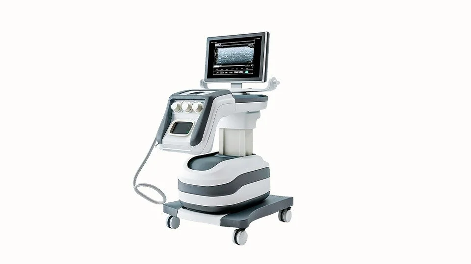
Physiological Vascular Testing is a non-invasive evaluation focused on assessing the physiological severity of the peripheral vascular pathology. The physiological testing serves as fast screening prior to deciding Imaging is required and provides different sets of required clinical input to decide immediate clinical intervention.(In contrast to Ultrasound Imaging or Duplex systems provide information about the anatomical severity and location of the pathology.)
Screening is easy, quick, and non-invasive, which aids in early diagnosis and advising early treatment, usually consisting of Medication and lifestyle changes.
In our Vein and foot Clinic we offer these Non Invasive Vascular tests as tool for clear insights into the health of the vascular system.
“In conclusion, We believe that early speciality referral and evaluation for any patient that has a concern for Vascular disease could represent a simple target that improves long-term outcomes and limits resource waste—ultimately, non-salvageable limbs.”.
Non Invasive Vascular lab
PURPOSE
Non-invasive physiologic evaluations are performed to determine the presence, severity, and general location of peripheral arterial occlusive disease (PAD). Intended Use of these Practice guidelines provide recommendations applicable to patients with or at risk of developing cardiovascular disease.
- Previous cardiovascular surgeries
- Current medications or therapies
- Presence of risk factors for peripheral arterial disease
- Diabetes
- Hypertension
- Hyperlipidemia
- Age
- Smoking
- Obesity
- Cerebrovascular disease
- Coronary artery disease
- Family history of PAD
- Presence of symptoms for peripheral Arterial Disease
Segmental Blood Pressures
Segmental blood pressures is a physiologic test which is performed using a physiologic machine in order to help in localizing arterial obstruction to flow along the limb as well as the physiological severity of the obstruction. This is in contrast to ultrasound imaging of the lower limb, which is focused on detecting and quantifying the anatomical severity of the arterial obstruction to flow.
Expected Results of Segmental Blood Pressures
When measuring segmental blood pressures, the focus is on the pressure difference between each 2 consecutive adjacent sites, as well as the pressure difference between similar sites on the right and left legs. For example, a large pressure drop between the thigh and the adjacent above-knee site suggests an arterial obstruction to flow, which is located between the thigh and the knee.
Ankle Brachial Index
What is Ankle Brachial Index (ABI)?
The Ankle Brachial Index is a standard and common method of physiologic assessment of vascular pathology in the lower limbs in a fast and simple manner. Ankle Brachial Index (ABI) test assists in the early detection of Peripheral Arterial Disease (PAD) and provides information about its functional severity. During the ABI test, the primary Systolic pressures are measured in each of the right and left brachials, as well as at the level of the right and left ankles.

Common Indications Often Used for an ABI Test
Intermittent claudication: Symptoms like leg pain, cramping, or numbness experienced during exercise
Non-healing wounds, post-operative Settings, Peripheral Arterial Disease Symptoms.
Toe Brachial Index (TBI)
What is Toe Brachial Index?
The Toe Brachial Index (TBI) is defined as the ratio between the systolic blood pressure in the right or left toe and the higher of the systolic pressure in the right or left arms.
TBI is a common vascular physiologic assessment test taken to determine the existence and severity of peripheral arterial disease (PAD) in the lower extremities.

Photo-Plethysmography
What is Photo-Plethysmography?
Photo-plethysmography (PPG) is a non-invasive testing method and is an essential tool in every physiological vascular testing examination.
The PPG sensors transmit infra-red waveforms to the skin and detect the signals that are reflected back to the sensor from the skin. The skin partially absorbs the transmitted signals, and the reflected signals are a function of this light absorption, which in turn is a function of the local blood perfusion. Therefore, the PPG waveforms reflect local and relatively shallow skin variations in blood flow.
Pulse Wave Velocity
What is Pulse Wave Velocity?
Pulse Wave Velocity (PWV) is a simple and non-invasive measurement which can be measured at various locations along with the arterial circulation to assess arterial stiffness.
Pulse wave velocity (PWV) is widely recognized as a simple and reliable clinical measure of arterial stiffness and elasticity, which is correlated with vascular disease. The contractions of the heart, which drive the arterial blood, also generate arterial blood pressure pulse waves, which propagate through the arterial walls. PWV is defined as the velocity at which these arterial blood pressure pulses propagate.
Older patients are expected to have more stiff arteries and, as a result, higher PWV values under normal conditions. Diabetic patients are also expected to have a significant increase in PWV values.
Non Invasive Venous lab
Venous Reflux Diagnosis; Assessing Venous Valve Competency in the Lower Legs
What is Venous Reflux?
The Venous Reflux test is used to determine the competence of the superficial venous valves in the calves of the legs. This test is performed with a DC PPG sensor.
When venous valves do not function properly, they may “leak,” causing blood flow in the veins to reverse. This, in turn, causes improper venous blood flow and blood pooling in the veins of the legs, which can lead to a variety of venous insufficiency diseases.
The Venous Reflux test is a common non-invasive diagnostic examination performed with a Phot plethysmography (PPG) sensor. It can be used to determine the competence of the venous valves in the lower legs.
Who Needs a Venous Reflux Test?
When patients experience symptoms like leg pain, heaviness, swelling, varicose veins, or changes in their skin, the Venous Reflux test may be necessary. These symptoms might point to a potential venous valve dysfunction.
In addition, the Venous Reflux test assesses chronic venous insufficiency, evaluates treatment effectiveness, aids preoperative evaluation, and monitors postoperative recovery in patients with venous disorders.
In some cases, the test may not be recommended or reliable. These cases include patients with Active deep vein thrombosis (DVT), Acute Inflammatory Conditions, Skin Integrity Issues, and allergic reactions to the adhesive used to attach the PPG sensor. in such cases, consider alternative methods or materials.
A targeted diagnostic evaluation of Venous Reflux test helps healthcare professionals make informed treatment decisions, improving patient care and outcomes.
Air Plethysmography
What is Air Plethysmography?
The Air Plethysmography (APG) test relates to the use of a pressure cuff as a sensor. The pressure cuff is inflated to low pressure, such as around 20 mmHg for lymphatic occlusion or around 60 mmHg for venous occlusion, and then the air displacement is used to measure blood volume changes at the measuring site.
Most clinical uses of APG are for diagnosis of venous disease and global lower limb hemodynamics, including chronic venous obstruction to flow, venous reflux, calf muscle pump action, or venous hypertension.
Don’t let vein issues affect your life.
Book a call with us
Maximum Venous Outflow / Segmental Venous Capacitance (MVO/SVC)
What is MVO/SVC?
The MVO/SVC test (Maximal Venous Outflow / Segmental Venous Capacitance) is a specific functional physiological lower limb venous test that helps diagnose obstructions to the venous circulation in the legs.
MVO/SVC
This test helps healthcare professionals assess the venous blood flow and capacitance ability of the venous circulation, which can indicate the presence of conditions like edema or other venous outflow obstructions.
If there is an obstruction to venous outflow, such as severe edema, the venous emptying from the plateau level will take longer. As a result, MVO will decrease, and the MVO/SVC ratio will also decrease compared to normal values.
Conditions We Treat
- Varicose veins
- Superficial Venous Thrombosis Phlebitis
- Thread / spider veins
- Swollen leg
- Deep vein thrombosis (DVT)
- May turners syndrome
- Varicose veins in pregnancy
- Varicose veins in obese patients
- Restless leg syndrome
- Varicose veins of the testicles
- Recurrent Varicose Veins
- Hidden Varicose Veins
- Hemosiderin Brown Stains
- Lipo dermatosclerosis (LDS)
- Venous Eczema
- Leg Ulcers
- Vaginal and Vulval Varicose Veins
- Pelvic Congestion Syndrome (PCS)

