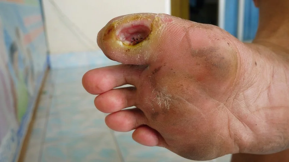
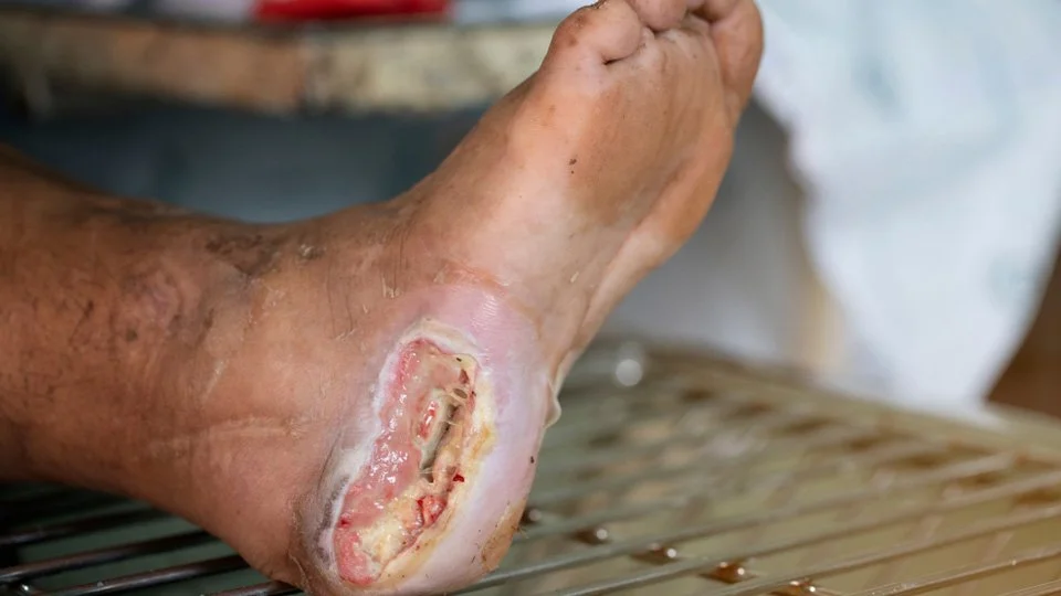
Diabetic foot Ulcers
Diabetic foot Ulcers
Diabetic foot Ulcers of the lower limbs are a major public health problem for which management requires further improvement, particularly in terms of healing time, prevalence, and recurrence rate.
The life time risk of a person with Diabetes having a foot ulcer could be as high as 25%.
Peripheral artery disease (PAD) of the lower extremities is a Diabetic rises to 55% in those above 80 years old. 15% of those with Diabetes for a decade suffer from Diabetic foot, which increases further to almost 50% by another decade.


Although most patients with PAD are asymptomatic, the disease increases the risk for cardiovascular morbidity and symptomatic disease progression. Patient prognosis after PAD diagnosis is poor because the disease often progresses to the extent that distal perfusion is insufficient to meet metabolic demands. This advanced PAD is commonly described as critical limb ischemia (CLI) and represents the end stage of the disease, mostly characterized by occlusive disease of the tibial and foot arteries in which patients suffer from rest pain, ischemic ulceration, and/or gangrenous tissue loss.
Arterial ulcers are typically more painful; affect the toes, heel, malleoli, or anterior shin; and are caused by arterial insufficiency.
Vein and foot clinic advocate a comprehensive plan of action to address all aspects of Critical limb ischemia in Diabetic foot ulcer, including diagnosis, treatment, and education of patients to control deadly impact of this disease.
DFU
The risk for developing CLI is considerably higher in diabetic patients and are more frequently reported in these patients compared to the general atherosclerotic population.
In diabetic patients, the immune system is impaired, so infection may be present without obvious signs.
Peripheral neuropathy generates 45% to 65% of DFS neuropathic or neuroischemic ulcers, and that patients with neuropathy express >3.5-fold higher risk for iterative foot ulceration. Ischemic ulcers are often described as “spontaneous” inferior limb ulcerations (typically located on the forefoot and toes) that occur when CIRCULATION to leg is severely compromised.
Arterial ulcers can also appear as “post-minor traumatic” wounds (e.g., common skin tears, cuts, blisters, abrasions, etc.) because local flow proves insufficient for ulcer to heal.
Don’t let vein issues affect your life.
Book a call with us
Although arterial ulcers can occur nearly anywhere on the ischemic limb, they are often found on the distal leg, toes, forefoot, or around the heel.
Arterial ulcers are generally associated with macrocirculatory peripheral artery disease (PAD) and are detected in 18% to 29% of people aged ≥ 60 years.
Advancements in technology are helping us achieve success in peripheral vascular interventional cases that were previously deemed impossible.
certain steps are taken to promote healing of an arterial-insufficient CLI wound. First, transcutaneous oximetry is performed in all patients. A number of studies have shown that periwound transcutaneous oxygen tension (PtcO2) below a cutoff of 40 mm Hg is associated with impaired healing due to inadequate oxygen supply.7-9 Thus, a PtcO2 < 40 mm Hg should prompt a referral to an invasive vascular specialist for possible revascularization.
In the ideal system of care, the wound should be treated with both an “outside-in” and an “inside-out” approach.
A wound care specialist takes care to use debridement, topical antimicrobials, systemic antibiotics, and HBO therapy to form the “outside-in” approach.
“inside-out” approach to restoring blood flow to the affected wound by the Vascular Surgeon. The goal of revascularization (endovascular intervention or vascular bypass surgery) is to facilitate ulcer healing.
Conditions We Treat
- Varicose veins
- Superficial Venous Thrombosis Phlebitis
- Thread / spider veins
- Swollen leg
- Deep vein thrombosis (DVT)
- May turners syndrome
- Varicose veins in pregnancy
- Varicose veins in obese patients
- Restless leg syndrome
- Varicose veins of the testicles
- Recurrent Varicose Veins
- Hidden Varicose Veins
- Hemosiderin Brown Stains
- Lipo dermatosclerosis (LDS)
- Venous Eczema
- Leg Ulcers
- Vaginal and Vulval Varicose Veins
- Pelvic Congestion Syndrome (PCS)

